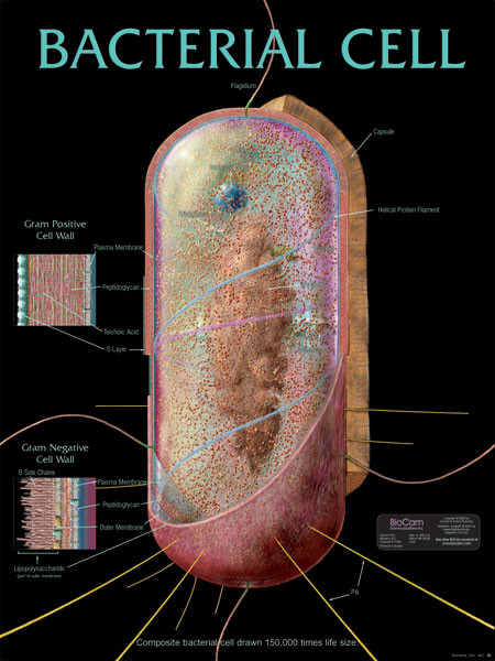
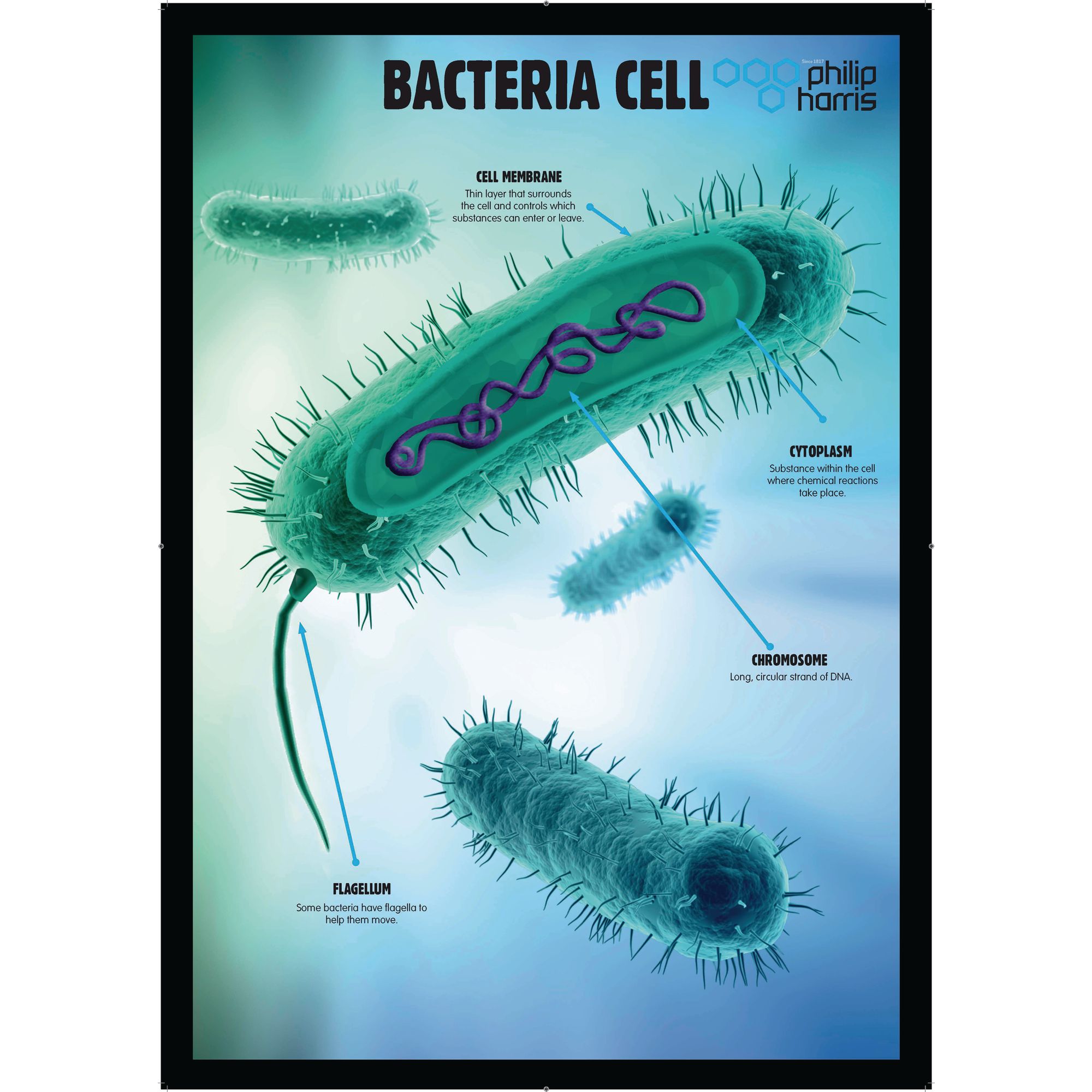
40 bacterial cell picture with labels
Bacterial Cell Structure Diagram - Quizlet Bacterial Cell Structure. Domain of unicellular prokaryotes that have cell walls containing peptidoglycan. an irregularly shaped region in a bacteria that contains all or most of the genetic information (either RNA or DNA). A nuclear membrane is not present. Hair like appendages found on the surface of most bacteria. Bacteria Cell Label - how to make a prokaryotic bacteria cell model ... Bacteria Cell Label. Here are a number of highest rated Bacteria Cell Label pictures on internet. We identified it from reliable source. Its submitted by running in the best field. We give a positive response this kind of Bacteria Cell Label graphic could possibly be the most trending subject ...
Bacterial cells - Cell structure - Edexcel - GCSE Combined Science ... Bacterial cells Bacteria are all single-celled. The cells are all prokaryotic. This means they do not have a nucleus or any other structures which are surrounded by membranes. Larger bacterial...
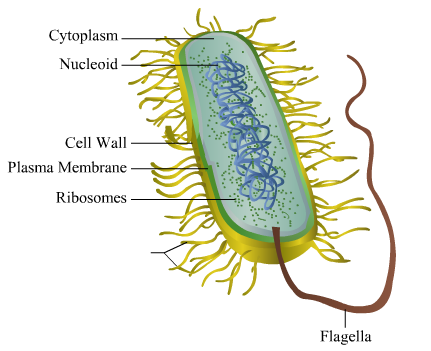
Bacterial cell picture with labels
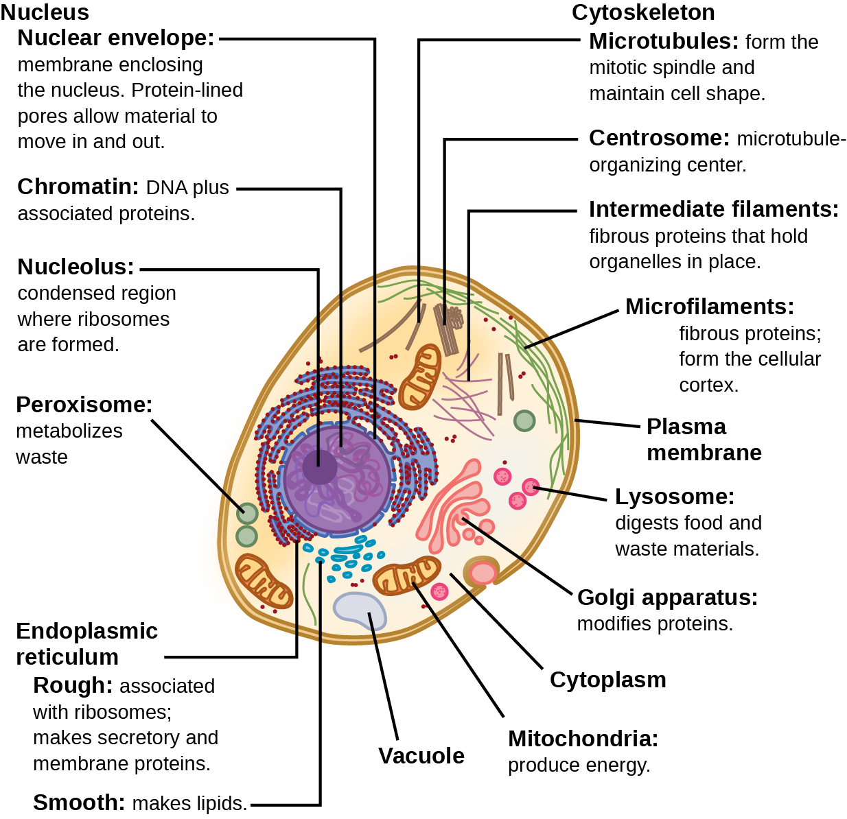
Animal Cell Labeled Pictures, Images and Stock Photos Browse 116 animal cell labeled stock photos and images available, or start a new search to explore more stock photos and images. Newest results Components of Eukaryotic cell, nucleus and organelles and plasma... Diagrams of animal and plant cells Components of Eukaryotic cell, nucleus and organelles and plasma... Honey labels. Bacteria - Definition, Structure, Diagram, Classification The bacteria diagram given below represents the structure of bacteria with its different parts. The cell wall, plasmid, cytoplasm and flagella are clearly marked in the diagram. Bacteria Diagram representing the Structure of Bacteria Structure of Bacteria The structure of bacteria is known for its simple body design. Bacteria (Prokaryote) Cell Coloring - The Biology Corner Color the cell wall purple. 2. On the inside of the cell wall is the cell membrane . Its job is to regulate what comes in and out of the cell. Color the cell membrane pink. 3. The surface of some bacteria cells is covered in pilus, which help the cell stick to surfaces. Color the pilus light green. 4.
Bacterial cell picture with labels. Draw a labelled diagram of a bacterial cell. - Careers360 The schematic diagram of bacterial cell structure. | Download ... Similar Questions. An Unbiased coin is tossed 4 times. What ...1 answer · Top answer: Draw a labelled diagram of a bacterial cell. A Labeled Diagram of the Animal Cell and its Organelles A Labeled Diagram of the Animal Cell and its Organelles There are two types of cells - Prokaryotic and Eucaryotic. Eukaryotic cells are larger, more complex, and have evolved more recently than prokaryotes. Where, prokaryotes are just bacteria and archaea, eukaryotes are literally everything else. 1. Draw a diagram of bacterial cell and label its parts. Classify ... Classify the bacteria according to its shape and give examples for each. Write in detail about Bacterial Capsule and Cell wall. II. Write notes on: (4 x 5 = 20).1 page 50 Striking Microscopic Images of Viruses and Bacteria Bacteria and viruses are some of the last things you hope to encounter on a day-to-day basis. After all, they're what make you sick. But if you look at bacteria, viruses and the cells they infect ...
Bacteria Labeled Diagram Stock Vector Image & Art - Alamy Download this stock vector: Bacteria Labeled Diagram - EG0XT7 from Alamy's library of millions of high resolution stock photos, illustrations and vectors. 600+ Free Bacteria & Virus Images - Pixabay Bacteria and virus high resolution images. Find your perfect picture for your project. 639 Free images of Bacteria / 7 ‹ › ... Bacteria Labeled Stock Illustrations - Dreamstime Bacteria Labeled Stock Illustrations - 220 Bacteria Labeled Stock Illustrations, Vectors & Clipart - Dreamstime Bacteria Labeled Illustrations & Vectors Most relevant Best selling Latest uploads Within Results People Pricing License Media Properties More Safe Search 220 bacteria labeled illustrations & vectors are available royalty-free. Next page Plant and Animal Cells - Labeled Graphics Other Cell Resources. Cheek Cell Lab - observe cheek cells under the microscope Cheek Cell Virtual Lab - if you missed it in class Animal Cell Coloring - color a typical animal cell. Plant Cell Coloring - color a typical plant cell Plant Cell Lab - microscope observation of onion and elodea Plant Cell Lab Makeup - microscope observation of onion and elodea, if students missed the lab that ...
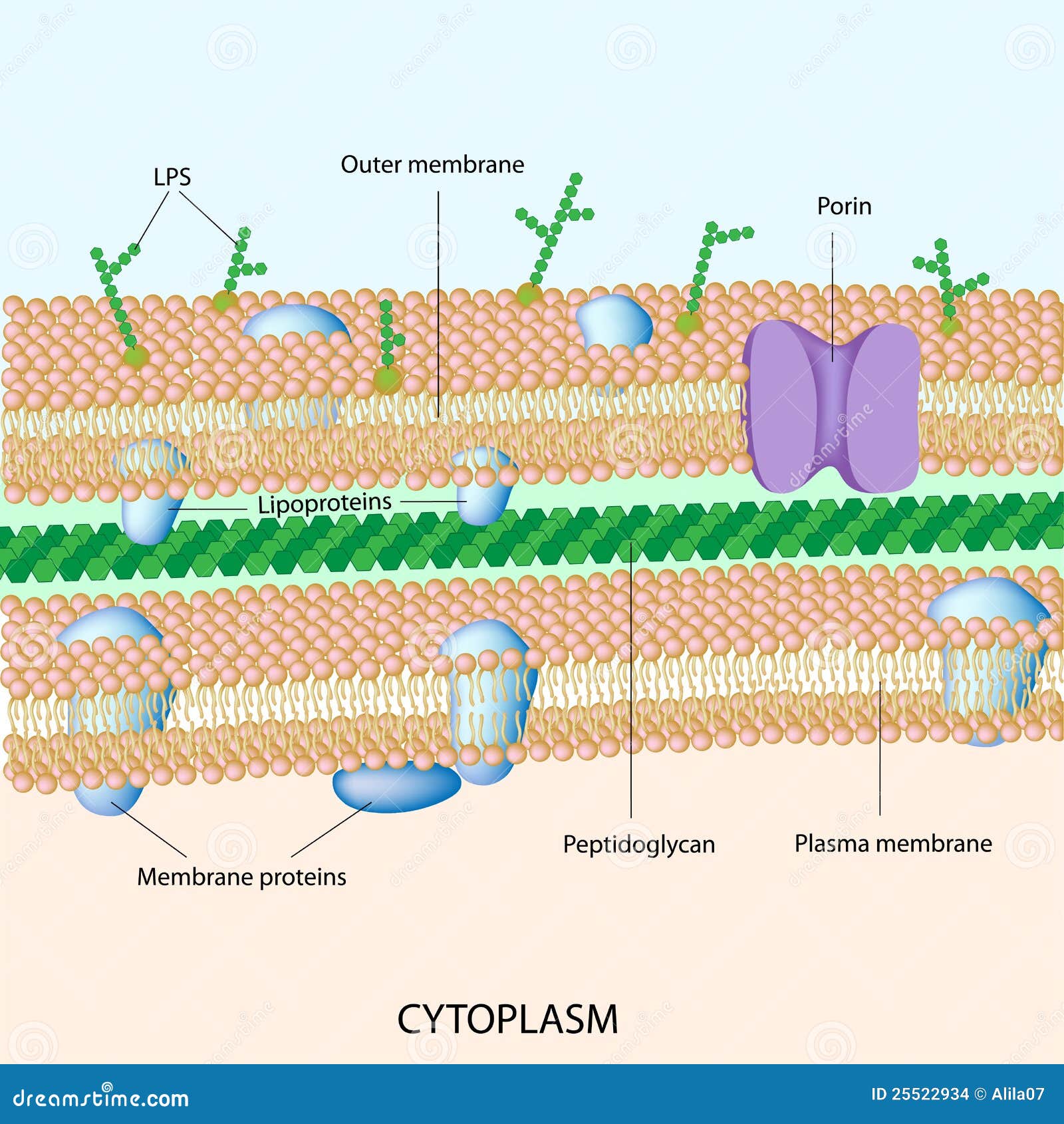
Structure of Bacteria (With Diagram) | Microbiology Bacterial cell wall is extremely thin (10-25 nm thick) and provides rigidity and a definite shape to the cell. 7. Chemically, the cell wall is composed of mucopeptide (murein) scaffolding or platform formed by N- acetyl glucosamine and N-acetyl muramic acid molecules arranged in alternate chains. 8. Structure of Bacterial Cell (With Diagram) - Biology Discussion It is a tough and rigid structure of peptidoglycan with accessory specific materials (e.g. LPS, teichoic acid etc.) surrounding the bacterium like a shell and lies external to the cytoplasmic membrane. It is 10-25 nm in thickness. It gives shape to the cell. Nucleus: The single circular double-stranded chromosome is the bacterial genome. Bacteria shapes, structure and diagram - Jotscroll Bacterial endospores layers Bacteria cells are the smallest living cells that are known; even though viruses are smaller than bacteria, viruses are not living cells. There are different types of bacteria with various sizes, shapes, and structures. The bacteria shapes, structure, and labeled diagrams are discussed below. Sizes Animal Cell Labeled Diagram Pictures, Images and Stock Photos Browse 19 animal cell labeled diagram stock photos and images available, or start a new search to explore more stock photos and images. Newest results Diagrams of animal and plant cells Labelled diagrams of typical animal and plant cells with editable layers. Golgi apparatus or Golgi body Lipids vector illustration.
en.wikipedia.org › wiki › Monoclonal_antibodyMonoclonal antibody - Wikipedia There may also be bacterial contamination and, as a result, endotoxins that are secreted by the bacteria. Depending on the complexity of the media required in cell culture and thus the contaminants, one or the other method (in vivo or in vitro) may be preferable. The sample is first conditioned, or prepared for purification.
› metronidazole › articleFlagyl (metronidazole) Side Effects, Pregnancy Use & Dosage Flagyl (metronidazole) is an antibiotic prescribed to treat various parasitic and bacterial infections (Giardia, C. diff, H. pylori). Common side effects are headaches, nausea, and metallic taste in the mouth. Pregnancy and breastfeeding safety information are provided.
3 Common Bacteria Shapes - ThoughtCo Bacteria Shapes The three basic shapes of bacteria include cocci (blue), bacilli (green), and spirochetes (red). PASIEKA/Science Photo Library/Getty Images By Regina Bailey Updated on August 20, 2019 Bacteria are single-celled, prokaryotic organisms that come in different shapes.
Different Size, Shape and Arrangement of Bacterial Cells Size of Bacterial Cell. The average diameter of spherical bacteria is 0.5-2.0 µm. For rod-shaped or filamentous bacteria, length is 1-10 µm and diameter is 0.25-1 .0 µm. E. coli , a bacillus of about average size is 1.1 to 1.5 µm wide by 2.0 to 6.0 µm long. Spirochaetes occasionally reach 500 µm in length and the cyanobacterium.
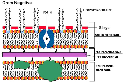
Label the image to demonstrate your understanding of bacterial cell ... Label the image to demonstrate your understanding of bacterial cell structures. Structure is differentiated by Gram stain procedure Provides mobility Often cany genes of antibiotic resistance and can be passed from one call to another Only found in gram-negative bacteria Common target for antibiotics due to a different structure from the eukaryotic version External structure for attachment to ...
Bacterial Staining Microbiology Images ... - Science Prof Online 1. Endospore stain of Bacillus subtilis showing both endospores (green) & vegetative cells (pink) @1000xTM; 2. Negative endospore stain showing only vegetative cells @1000xTM; 3. Malachite green primary staining step of endopore stain with slide being heated over water bath; 4. Applying counterstain (safrinin) to bacterial smear as last step of endospore stain; Endospore stained slide, with ...
PHOTO GALLERY OF BACTERIA - Microbiology in pictures Mixture of bacteria on agar plate. Bacterial colonies. Bacteria and pigment production. Bacterial colonies and hemolysis. Bacteria: Light microscopy. Bacteria.... Bacteria images. Colonies of various bacteria. Bacteria photos
Interactive Bacteria Cell Model - CELLS alive The three primary shapes in bacteria are coccus (spherical), bacillus (rod-shaped) and spirillum (spiral). Mycoplasma are bacteria that have no cell wall and therefore have no definite shape. Outer Membrane: This lipid bilayer is found in Gram negative bacteria and is the location of lipopolysaccharide (LPS) in these bacteria.
Image Library | CDC Online Newsroom | CDC Under a high magnification of 21674X, this digitally-colorized, scanning electron microscopic (SEM) image depicts a view of a dividing, Escherichia coli bacterium, clearly displaying the point at which the bacteria's cell wall was splitting into two separate organisms. See PHIL 7137 for a black and white version of this image.
Bacteria Cell Structures with Labels Stock Vector - Dreamstime Get 15 images free trial Bacteria Cell Structures with labels Royalty-Free Vector Bacterial cell structures labeled on a bacillus cell with nucleoid DNA and ribosomes. External structures include the capsule, pili, and flagellum. Morphology of internal structures of bacteria. cell anatomy bacteria, prokaryotic cell, cell, internal structures,
how to draw & label bacteria - YouTube | Science diagrams, Teaching, Labels Illustration of Set of bacterium and microorganism vector art, clipart and stock vectors. Image 20169845. Find support stock images in HD and millions of other royalty-free stock photos, illustrations and vectors in the Shutterstock collection. Thousands of new, high-quality pictures added every day.
› articles › nmethPooled CRISPR screening with single-cell transcriptome ... Jan 18, 2017 · Pooled CRISPR screening is a powerful and widely used method for identifying genes involved in biological mechanisms such as cell proliferation 1,2, drug resistance 3, and viral infection 4.Cells ...
97,783 Bacteria Cell Stock Photos and Images - 123RF Bacteria Cell Stock Photos And Images 97,783 matches Page of 978 Structure of a bacterial cell. Anatomy of the prokaryote. unicellular organism. Vector diagram for your design, educational, medical, biological and science use Bacteria vector icon isolated on transparent background, Bacteria logo concept
plantvillage.psu.edu › topics › pepper-bellPepper, bell | Diseases and Pests, Description, Uses, Propagation Initial symptoms of infection are the formation of small, circular, water-soaked spots on leaves, stems, petioles and/or peduncles; the lesions mature to have white to brown centers surrounded by a brown to red or purple border; as the lesions expand, they may develop a water-soaked outer edge and dark outer ring which gives the lesions a concentric appearance; mature lesions are brittle and ...
Label the Bacterium Cell - EnchantedLearning.com Label the Bacterium Cell - EnchantedLearning.com The cell is the basic unit of life. The following is a glossary of Bacterium cell terms. basal body - A structure that anchors the base of the flagellum and allows it to rotate. capsule - A layer on the outside of the cell wall. Most but not all bacteria have a capsule.
Structure of a bacterial cell, labeled. Stock Illustration Download Structure of a bacterial cell, labeled. Stock Illustration and explore similar illustrations at Adobe Stock. Adobe Stock. Photos Illustrations Vectors Videos Audio Templates Free Premium Editorial Fonts. Plugins. 3D. Photos Illustrations Vectors Videos Audio Templates Free Premium Editorial Fonts.
Bacteria (Prokaryote) Cell Coloring - The Biology Corner Color the cell wall purple. 2. On the inside of the cell wall is the cell membrane . Its job is to regulate what comes in and out of the cell. Color the cell membrane pink. 3. The surface of some bacteria cells is covered in pilus, which help the cell stick to surfaces. Color the pilus light green. 4.




Post a Comment for "40 bacterial cell picture with labels"